0:29, silent. Watch it with music on Instagram.
A 7 Tesla brain MRI, slice by slice
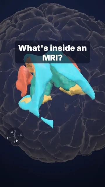
The same 7 Tesla brain, scrolled through in coronal slices, with its deep structures pulled out in 3D.
Most hospital MRI scanners run at 1.5 or 3 Tesla. This scan was made at 7 Tesla, with 0.25 mm voxels: each one has 1/64 of the volume of a standard 1 mm voxel. This time we scroll through the same brain in coronal slices, from the back of the thalamus to the front of the basal ganglia, and pull out its deep structures in 3D:
- caudate nucleus
- putamen + 🩷 globus pallidus = the lentiform nucleus, named for its lens shape
- thalamus: relays almost all sensory input to the cortex
Sources and licence
Open data: Lüsebrink et al., Sci Data 2021 (OpenNeuro ds003563; CC0). 3D models segmented from the same volunteer with FreeSurfer; globus pallidus divided with MASSP 2.0 (Bazin et al. 2025, CC BY 4.0).
Video licence: CC BY 4.0. To reuse it, credit “Cerebrarium (Aldo Spolaore)”, link to this page and name the data sources above.
Educational content, not medical advice.