0:33, silent. Watch it with music on Instagram.
What's hiding in a brain MRI
What are you actually looking at on a brain MRI?
A 7 Tesla MRI, slice by slice, part 1 of 2Next: A 7 Tesla brain MRI, slice by slice
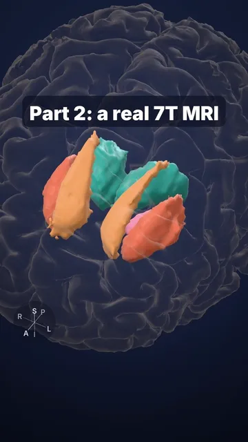
This is a real T1-weighted 7 Tesla scan at 0.25 mm resolution. On T1, fluid is dark, white matter is bright and grey matter is in between. As the slice moves down, the deep structures of this same brain rise out in 3D:
- lateral ventricles: CSF-filled cavities
- caudate nucleus + putamen: the (dorsal) striatum, which helps select and start movements and forms habits
- thalamus: relays almost all sensory input to the cortex
- hippocampus: forms new memories of facts and events
Sources and licence
Open data: Lüsebrink et al., "Comprehensive ultrahigh resolution whole brain in vivo MRI dataset as a human phantom", Sci Data 2021 (doi:10.24352/ub.ovgu-2020-145, mirrored on OpenNeuro ds003563; CC0). 3D models segmented from the same volunteer with FreeSurfer.
Educational content, not medical advice.