0:31, silent. Watch it with music on Instagram.
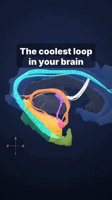
200 million fibres: the corpus callosum
A real 7 Tesla brain MRI: the bright arc is the corpus callosum, about 200 million nerve fibres joining the two halves of the brain
Most hospital MRI scanners run at 1.5 or 3 Tesla; this volunteer was scanned at 7 Tesla. On a sagittal MRI the corpus callosum is the bright arc above the ventricles. Its front part (genu) links the frontal lobes, and its back part (splenium) links the occipital and temporal lobes.
In some severe epilepsies that drugs can't control, surgeons cut it (corpus callosotomy) to stop seizures spreading. In lab tests, a split-brain patient can't name an object flashed to the left visual field. The right hemisphere sees it, but speech sits in the left hemisphere in most people. Yet the left hand, run by the right hemisphere, can still pick the object out by touch.
Sources and licence
Rendered from open data: lobes, motor strip and 7 T MRI of a volunteer (Lüsebrink et al., Sci Data 2021, CC0); callosal fibres from the HCP1065 atlas (Yeh, Nat Commun 2022, CC BY-SA 4.0), data provided by the Human Connectome Project, WU-Minn Consortium (Principal Investigators: David Van Essen and Kamil Ugurbil; 1U54MH091657) funded by the 16 NIH Institutes and Centers that support the NIH Blueprint for Neuroscience Research; and by the McDonnell Center for Systems Neuroscience at Washington University. Hemispheres pulled apart and fibre colours drawn schematically.
Video licence: CC BY-SA 4.0. To reuse it, credit “Cerebrarium (Aldo Spolaore)”, link to this page and name the data sources above.
Educational content, not medical advice.