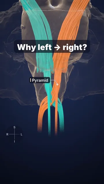
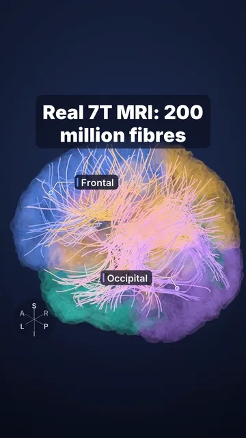
0:32, silent. Watch it with music on Instagram.
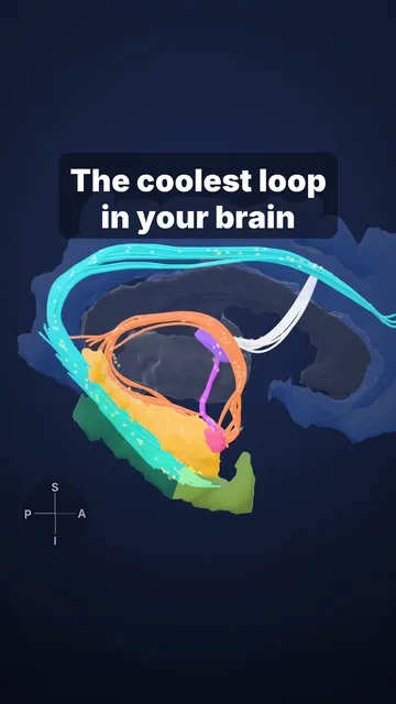
Vision takes a detour: Meyer's loop
Why can a lesion in the temporal lobe cause a visual field defect?
Visual pathway: optic nerves → optic chiasm (fibres from the nasal half of each retina cross) → optic tracts → lateral geniculate body of the thalamus → optic radiation → primary visual cortex along the calcarine sulcus.
The front bundle of the optic radiation, Meyer's loop, runs forward over and lateral to the temporal horn of the lateral ventricle before turning back. It carries the opposite upper visual field (from the lower retina). Damage to it (for example from a tumour or an anterior temporal lobectomy) causes a contralateral homonymous superior quadrantanopia: the upper quarter of vision on the opposite side is lost.
Sources and licence
Rendered from open data: 7 T MRI and brain of a volunteer (Lüsebrink et al., Sci Data 2021, CC0), with optic nerves, chiasm and tracts traced by us on the same scan; lateral geniculate bodies from MASSP 2.0 (Bazin et al. 2025, CC BY 4.0); optic radiations from the HCP1065 atlas (Yeh, Nat Commun 2022, CC BY-SA 4.0), data provided by the Human Connectome Project, WU-Minn Consortium (Principal Investigators: David Van Essen and Kamil Ugurbil; 1U54MH091657) funded by the 16 NIH Institutes and Centers that support the NIH Blueprint for Neuroscience Research; and by the McDonnell Center for Systems Neuroscience at Washington University. Crossing fibres, eyes and visual fields drawn schematically.
Video licence: CC BY-SA 4.0. To reuse it, credit “Cerebrarium (Aldo Spolaore)”, link to this page and name the data sources above.
Educational content, not medical advice.