0:39, silent. Watch it with music on Instagram.
Name 5 deep brain structures
A quiz on five deep structures of one real brain, shown on its 7 Tesla MRI.
Name it quiz, part 1 of 3Next: Which artery feeds each region?
Pause the video at each structure and name it before you open the answers.
Show the answers
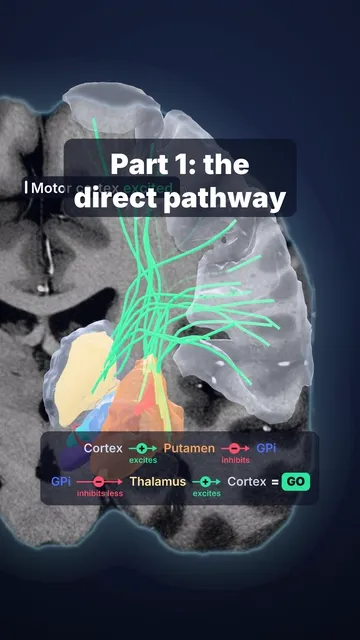
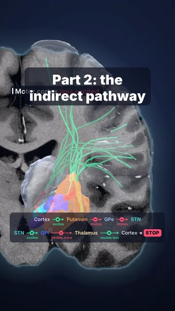
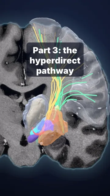
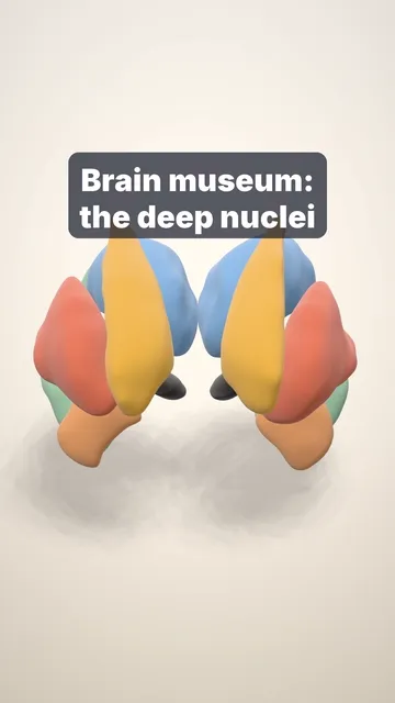
- Caudate nucleus: its head bulges into the frontal horn of the lateral ventricle. In Huntington's disease it wastes away and the horn widens.
- Thalamus: every sense except smell is relayed here on its way to the cortex.
- Putamen: the most common site of a hypertensive brain bleed.
- Globus pallidus: its inner part (GPi) is a deep brain stimulation target in Parkinson's disease and dystonia.
- Hippocampus: key for forming new memories. It shrinks in Alzheimer's disease.
Sources and licence
Data: 7 T MRI of one volunteer and its 3D segmentation (Lüsebrink et al., Sci Data 2021, CC0), smoothed for display; the highlighted regions are the cross-sections of those models on the same scan. Facts: Radiopaedia (Huntington disease; hypertensive intracerebral haemorrhage; Alzheimer disease); StatPearls, Neuroanatomy, Thalamus; Okun, N Engl J Med 2012 (deep brain stimulation).
Video licence: CC BY 4.0. To reuse it, credit “Cerebrarium (Aldo Spolaore)”, link to this page and name the data sources above.
Educational content, not medical advice.